Leonardo da Vinci anatomical drawings stand as a testament to his genius, marrying art and science in unprecedented ways.
These drawings showcase his artistic skill and deep interest in understanding the human body.
Leonardo’s work, created when scientific illustration was rare, provided detailed insights into human anatomy ahead of its time.
Leonardo’s work offers a perfect example for those curious about how art and science intersect.
His anatomy studies began as part of his artistic endeavors but evolved into something more significant. His ability to dissect and accurately depict the human form was artistic and scientific.
Exploring Leonardo’s anatomical sketches transports one into his world. There, he dissected bodies, often at night, by candlelight, with cloth covering his mouth and nose.
These drawings were part of his ambitious project to create an anatomical treatise, which was unfortunately lost for centuries. Nevertheless, they remain a significant contribution to art and science today.
Leonardo da Vinci: A Brief Biography
Leonardo da Vinci was born in Vinci, Italy, on April 15, 1452. As a polymath of the Renaissance, he excelled in various fields, such as art, science, and engineering. His artistic masterpieces, including the “Mona Lisa” and “The Last Supper,” are celebrated worldwide.
He was not only skilled in painting but also created intricate anatomical drawings.
These sketches demonstrated a remarkable understanding of the human body. His observations contributed significantly to both art and science.
In his lifetime, Leonardo produced numerous anatomical sketches that displayed his keen eye for detail.
Leonardo’s interest in anatomy led him to dissect human bodies. He made extensive notes and drawings that revealed the structure of muscles, bones, and organs.
These insights are considered groundbreaking in the field of human anatomy even to this day.
Besides being an anatomist, Leonardo was also an inventor. His sketches of flying machines, war engines, and other devices highlight his inventive mind.
Although many of his inventions were never built, they paved the way for future innovations.
Leonardo’s talents extended far beyond anatomy and art. He was also a skilled musician, architect, and mathematician. His diverse abilities made him a true Renaissance man.
Overview of da Vinci’s Anatomical Studies
Leonardo da Vinci’s anatomical studies fused art and science, advancing our understanding of the human body. His work included detailed anatomical drawings and observations, highlighting the potential of art to convey complex scientific ideas.
Historical Context
During the Renaissance, the focus on humanism and the pursuit of knowledge fostered a new interest in understanding the human body.
Leonardo da Vinci thrived in this vibrant intellectual environment, influenced by contemporaries like Leon Battista Alberti. Alberti encouraged artists to depict human figures based on anatomy.
Da Vinci started his anatomical studies in the late 15th century, during his time in Milan. A blend of traditional beliefs and direct observations from dissections influenced these studies.
His methodical approach and detailed illustrations set his work apart from previous studies.
The anatomy drawings da Vinci created remained superior in accuracy and artistic expression, illustrating muscles, bones, and organs in ways that had never been seen before.
His collaboration with doctors and access to dissection resources significantly contributed to his understanding and detailed sketches.
Major Contributions to Anatomy
Leonardo da Vinci anatomical drawings, particularly his studies of the human skeleton, muscles, and organs, marked a significant advancement in the field.
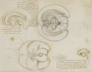
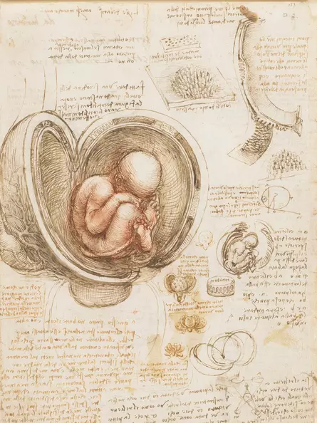
He produced pioneering studies of the human brain, heart, and prenatal development.
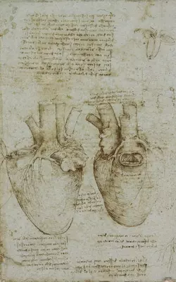
His work revealed groundbreaking insights, such as the accurate depiction of the heart’s ventricles and the function of the aortic valve, years before they were officially recognized.
Many of his discoveries were documented in meticulous drawings and notes, intended for a comprehensive anatomy book that was never published in his lifetime.
The Renaissance master’s blending of precise artistic techniques with anatomical research, exemplified in Leonardo’s Study of Anatomy, helped set a foundation for modern anatomy.
Techniques and Materials Used
Leonardo da Vinci anatomical drawings are renowned for their precision and detail. Leonardo set new standards in studying human anatomy by combining artistic skill with scientific inquiry.
His choice of methods and materials was crucial in these groundbreaking works.
Drawing and Dissection Methods
Leonardo systematically blended artistic techniques with scientific methods. He often conducted dissections to gain firsthand insight into human anatomy.
This hands-on approach allowed him to create realistic anatomical sketches grounded in observation.
By carefully examining muscles, bones, and organs, Leonardo depicted the human body with unparalleled accuracy, which some studies describe as akin to viewing an anatomy textbook.
His meticulous dissection practices and keen observation skills informed these works, ensuring his drawings were scientifically valuable and artistically compelling.
Paper and Ink Selection
Leonardo’s choice of materials was key in producing his detailed drawings.
He often used high-quality paper that could withstand his work’s fine lines and shading.
Ink, crafted from natural substances, provided the rich contrasts he needed for his chiaroscuro technique. This method, involving the interplay of light and dark, offered a sense of depth and realism in his anatomical sketches.
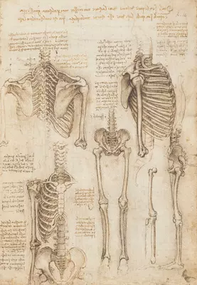
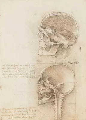
Many of his drawings, such as his studies on the human spine, remain influential, highlighting the importance of material selection in the longevity and impact of his art.
This strategic use of materials contributed significantly to the enduring brilliance of da Vinci’s anatomical studies.
The Vitruvian Man: Combining Art and Anatomy
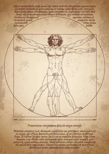
The Vitruvian Man by Leonardo da Vinci is a remarkable fusion of art and science. This iconic drawing features a nude male figure in two superimposed positions. The figure is perfectly inscribed in a circle and a square, highlighting proportions inspired by the work of the ancient Roman architect Vitruvius.
Leonardo, known for his profound anatomical drawings, used his talents to explore the human form deeply.
His studies in anatomy, reflected in the Vitruvian Man, demonstrate the intersection of geometry and human structure.
These observations emphasize how the human body reflects the order of the universe.
Da Vinci’s meticulous approach to anatomical sketches illustrates his dedication to understanding the intricacies of the body.
By dissecting and observing human bodies, he developed insights far ahead of his time. His work bridged the gap between artistic representation and scientific examination.
The Vitruvian Man is more than just a drawing; it is a visual embodiment of Renaissance humanism.
This perspective appreciates humans as the center of the universe while celebrating their connection with the world.
Da Vinci’s drawing recruits principles from art and science, representing his belief in the harmony between nature and man.
In addition to its artistic prowess, the piece symbolizes Leonardo da Vinci’s role as an anatomist.
His pursuit of knowledge through Leonardo da Vinci anatomy drawings set a precedent for future studies. By merging artistic skill with scientific inquiry, he brought the world a new understanding of both disciplines.
Human Anatomy
Leonardo da Vinci anatomical drawings provided valuable insights into the human body, focusing on its intricate structures and functions. These drawings highlight key areas such as the skeleton, musculature, and internal organs.
Skeleton
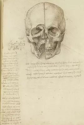
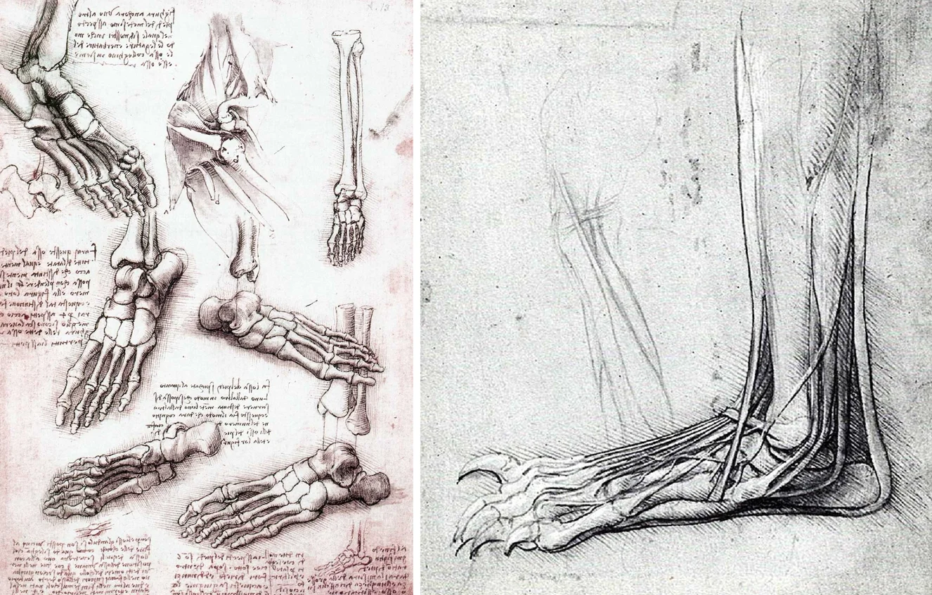
The skeleton is depicted with remarkable accuracy in da Vinci’s anatomical sketches.
He illustrated each bone’s position and connection within the human body.
His study of the skeletal system showed an understanding of its supportive structure, which underlies all bodily movements.
Joint mechanics, including how bones like the femur and humerus work together to provide motion, were given detailed attention.
Musculature
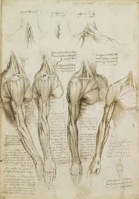
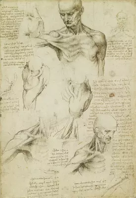
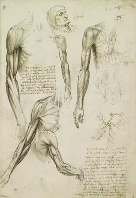
Leonardo da Vinci’s anatomy studies also displayed a profound examination of musculature.
He meticulously recorded the layers of muscles, highlighting their placement and function.
His drawings often show muscles in action, revealing the complex interplay that allows for varied movements.
Through this work, musculature was shown not just as static elements but as dynamic parts essential for the human body’s performance.
Internal Organs and Heart Muscle
Da Vinci’s exploration of internal organs was groundbreaking.
His studies included the positioning and function of organs within the body cavity.
The heart was a particular focus, as his drawings showcased its chambers and movement.
His work helped pave the way for modern cardiovascular understanding, visually capturing the heart muscle and circulation principles.
Blood Vessels
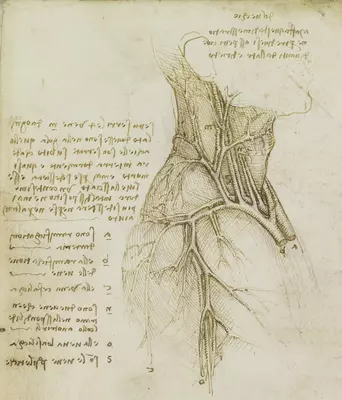
The depiction of blood vessels in da Vinci’s work provided insights into their pathways and connections.
He drew detailed maps of the circulatory system, noting how vessels supply blood across the body.
These anatomical drawings show the relationship between major arteries and veins, emphasizing the complexity of the circulatory network.
Nervous System
Leonardo’s study of the nervous system addressed the intricacies of neural pathways and brain structure.
His anatomical sketches depicted the distribution of nerves and their role in coordinating body functions.
Although his knowledge was limited by the scientific understanding of his time, his work showed keen interest in the relationship between the brain and the body.
Sexual Organs and Reproduction
Da Vinci’s investigations into sexual organs and reproduction demonstrated a systematic approach to understanding human generation.
His illustrations covered male and female reproductive anatomy, documenting their structures in detail.
In these studies, da Vinci aimed to convey the biological processes of reproduction, although his interpretations were influenced by his era’s cultural and medical limitations.
Animal Anatomy and Comparative Anatomy
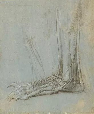
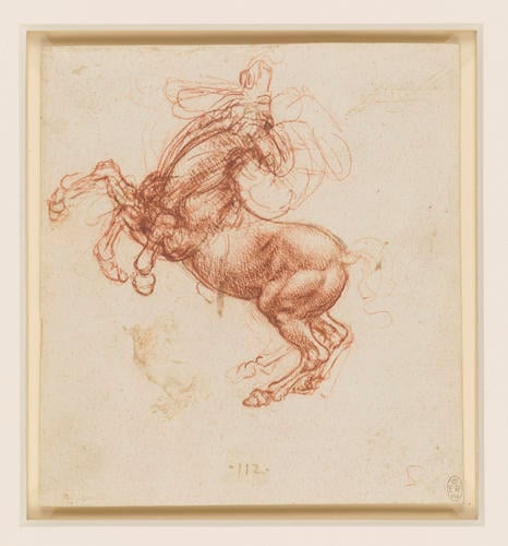
Leonardo da Vinci’s anatomical drawings showcased human anatomy and explored animal forms. For example, his studies of bears and horses testify to his deep curiosity about the similarities and differences between species. These works reveal his commitment to understanding the essence of life in all its forms.
Da Vinci meticulously observed how the anatomy of animals compared to humans. He noted shared features, such as muscles and skeleton structures, which he captured through detailed anatomical sketches. His ability to highlight these parallels underscores his expertise in both art and science.
Leonardo often focused on specific parts in these sketches, such as the limbs and joints. This focus helped him illustrate how the movement and strength of animals were similar yet distinct from those of humans. By comparing these aspects, da Vinci contributed valuable insights to comparative anatomy.
Leonardo da Vinci’s art techniques made complex details accessible. Bold lines, shading, and careful observation are evident in his work, providing depth and realism to his drawings. These techniques helped convey his findings in a visual, informative, and aesthetically pleasing form.
His animal anatomy studies influenced many fields, including medicine and biology. Today, his anatomical drawings remain valuable resources for those studying the links between human and animal physiology. His work inspires artists and scientists, bridging the gap between art and science.
Da Vinci’s Influence on Modern Medicine
Leonardo da Vinci’s anatomical drawings had a profound impact on the field of modern medicine. By pioneering new approaches to understanding the human body, da Vinci laid the groundwork for advancements in surgical techniques and medical education.
Surgical Techniques
Da Vinci’s anatomical sketches helped transform surgical practices. His detailed studies of the human form, including bones, muscles, and organs, allowed for a more precise and accurate understanding of human anatomy.
His medical drawings showed how organs functioned and fit into the body, offering insights critical for more effective surgical techniques.
Surgeons began employing more precise and informed methods, improving surgical outcomes. His work with dissecting cadavers revealed crucial insights into how surgeries could be performed more safely and efficiently.
This contributed significantly to the evolution of surgical instruments and techniques, many of which are still influenced by his findings today.
Educational Legacy
Leonardo da Vinci anatomical drawings are also vital to medical education. His illustrations were renowned for their clarity and detail, making them teaching tools for centuries. In his collaboration with Marcantonio della Torre at the University of Pavia, he created comprehensive anatomical sketches of the human body.
These drawings served as educational material, guiding medical students and practitioners in accurately identifying and understanding different bodily structures. Da Vinci’s ability to blend art with science allowed his anatomy manuals to convey complex information effectively.
His educational impacts resonate in medical schools today, where visual aids remain crucial for training future medical professionals.
Challenges and Controversies
Leonardo da Vinci’s anatomical drawings demonstrate his keen interest in understanding the human body. He faced many obstacles and criticisms.
Key issues included the Church’s resistance to dissections and questions about the accuracy of some of his sketches.
Church Opposition
The Church strongly influenced societal norms during Leonardo da Vinci’s time. Due to religious beliefs, the Church often opposed dissections of human bodies. Leonardo conducted many of his studies in secret to avoid controversy.
Despite this risk, his detailed anatomical sketches laid the groundwork for future science. His courage helped move scientific thinking forward, but his work faced limitations from the religious restrictions of his era.
Anatomical Inaccuracies
Although Leonardo’s drawings were groundbreaking, they contained some inaccuracies. This was partly due to the limited scientific knowledge of the time and restricted access to bodies for dissection.
Some of his drawings contained errors in organ placement or proportions. Despite these inaccuracies, his attempts to detail human anatomy were revolutionary. He prioritized understanding the human form with a precision that surpassed many of his contemporaries.
Preservation and Digitization of the Drawings
Leonardo da Vinci’s anatomical drawings have fascinated scholars and artists for centuries. His intricate human body sketches testify to his keen observations and artistic mastery. Preserving these masterpieces ensures they remain accessible for future generations.
Museums and galleries worldwide have taken steps to store and display Leonardo da Vinci’s anatomy drawings securely.
These institutions often use climate-controlled environments to maintain the integrity of the delicate paper and ink. Regular inspections ensure that any signs of deterioration are promptly addressed.
Digitization is crucial in preserving Leonardo da Vinci’s work. He converted his anatomical drawings into digital formats by scanning them at high resolution.
This protects the original pieces and allows people worldwide to explore his genius without needing to view them in person.
Interactive platforms make the experience even more prosperous. Online collections, like the Royal Collection Trust, provide detailed annotations and zoom features, allowing users to appreciate every stroke and detail of Leonardo da Vinci’s anatomy sketches.
These efforts continue Leonardo da Vinci’s legacy as a pioneering anatomist. Combining traditional conservation techniques with modern digital tools provides a comprehensive approach to preserving and sharing his invaluable medical drawings with a global audience.
Display and Exhibition of the Anatomical Works
Leonardo da Vinci anatomical drawings continue to fascinate the public. These sketches, which showcase his deep study of human anatomy, have been displayed in various renowned exhibitions. The Queen’s Gallery hosted one such exhibition, providing a rare chance to view these masterpieces.
Da Vinci’s studies involved meticulous dissection and careful observation. These pioneering sketches highlight his revolutionary approach, blending art with science.
Today, the Royal Collection Trust holds many of these works and occasionally displays them publicly, captivating audiences with their historical and scientific significance.
The exhibitions often pair da Vinci’s work with modern imagery, such as MRI scans, illustrating how his techniques foreshadowed today’s medical imaging. Visitors can see original 16th-century bindings in some events, adding a touch of history to their experience.
Curators emphasize the lasting impact of da Vinci’s innovative methods by displaying his drawings alongside contemporary anatomical images. These exhibitions allow people to appreciate his work’s artistic and scientific value.
Seeing Leonardo da Vinci’s anatomical sketches is a unique educational experience. It bridges historical achievements and modern understanding, offering insights into the early study of human anatomy and the genius behind these illustrations.
Final Thoughts
Leonardo da Vinci anatomical drawings are a remarkable blend of art and science. His work has profoundly influenced both fields, as he meticulously studied the human body to improve his art. These drawings remain significant, showcasing his genius and passion for understanding the human form.
Leonardo examined and sketched human anatomy while working alongside scholars like Marcantonio della Torre at universities. His techniques were ahead of his time, reflecting his dedication to accuracy and detail. His illustrations captured the intricacies of muscles, bones, and organs.
Leonardo’s use of dissection allowed him to observe the human body intimately. Despite the challenging conditions of his time, he created what would become some of the most precise anatomical works of the Renaissance. His sketches, like his study of the human spine, are still used in medical schools as reference material.
His works demonstrate a profound understanding of how art and anatomy intersected during his era. Examining his studies gives insight into his dual role as an artist and a scientist. These anatomical drawings not only informed his paintings but also paved the way for future studies in anatomy.
Frequently Asked Questions
Leonardo da Vinci significantly contributed to anatomical studies, illustrating the human body with remarkable detail. These drawings explored various aspects of human anatomy, from the heart to the muscular system.
Did Leonardo da Vinci make anatomical drawings?
Yes, Leonardo da Vinci created detailed anatomical drawings. These works are celebrated for their accuracy and depth, reflecting his interest in the human body. His drawings are still studied as vital historical contributions to anatomy.
Did Leonardo da Vinci draw the heart?
Leonardo da Vinci drew the heart, focusing on its complex structure. His depiction of the heart includes detailed observations that were advanced for his time. This work is housed in the Royal Collection Trust at Windsor Castle, England.
Why did Leonardo da Vinci draw skeletons?
He drew skeletons to understand the body’s framework. He believed that knowledge of bones would improve his artistic portrayal of the human form. This study was part of his broader exploration of anatomy during the Renaissance.
What is Leonardo da Vinci’s famous drawing?
Leonardo’s most famous drawing is the Vitruvian Man. According to the Roman architect Vitruvius, this drawing illustrates the ideal human proportions. It combines art and science to highlight human symmetry and proportion.
Is Leonardo da Vinci the father of anatomy?
Leonardo da Vinci significantly influenced anatomical study but is not considered the “father of anatomy.” Although his contributions provided valuable insights into human biology, this title often goes to other historical figures in the field.
Who is the father of anatomy?
Andreas Vesalius is widely considered the father of anatomy. In 1543, he authored De humani corporis fabrica, a groundbreaking book on human anatomy that laid the foundation for modern anatomical studies.
How did Leonardo da Vinci contribute to our understanding of the human muscular system?
Leonardo da Vinci contributed by illustrating various muscle groups in detail. His studies showed how muscles interact and function within the human body, and his drawings remain a valuable reference for understanding musculature.
Who is the greatest anatomist of all time?
Naming the greatest anatomist can be subjective. Andreas Vesalius is one of the most renowned for revolutionizing anatomical study. His detailed work on human dissection set new standards for accuracy and detail in the field.
Who painted the Vitruvian Man based on his study of human anatomy?
Leonardo da Vinci painted the Vitruvian Man, a depiction based on his study of human anatomy and proportions. The drawing exemplifies the blend of art and science during the Renaissance.
Where is Leonardo da Vinci buried?
Leonardo da Vinci is buried at the Chapel of Saint-Hubert in Amboise, France. He spent the final years at the Château du Clos Lucé, where his grave is in a small chapel on the estate’s grounds.
How did Michelangelo study anatomy?
Michelangelo studied anatomy through dissection. He examined the human body to enhance his sculptural and artistic works.
Like Leonardo, he combined anatomy knowledge with his art for more lifelike representations.
 I’m Leonardo Bianchi, the mind behind Leonardo da Vinci's Inventions. Thanks for visiting.